

 |  |  |
| | ||
Knee arthroscopy - series: Procedure, part 2
 |
|
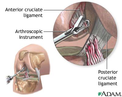
The viewing scope (arthroscope) and other instruments are inserted into the knee joint. The surgeon can see the ligaments, the knee disc (meniscus), the knee bone (patella), the lining of the joint (synovium), and the rest of the joint. Damaged tissues can be removed. Arthroscopy can also be used to help view the inside of the knee while ligaments or tendons are repaired from the outside.
Update Date: 5/12/2008 Updated by: Thomas N. Joseph, MD, Private Practice specializing in Orthopaedics, subspecialty Foot and Ankle, Camden Bone & Joint, Camden, SC. Review provided by VeriMed Healthcare Network. Also reviewed by David Zieve, MD, MHA, Medical Director, A.D.A.M., Inc.
